Described by Schmidle et Lauterborn in 1900! Lets first pause to appreciate that statement….Although it was Antony Van Leeuwenhoek who invented the microscope in the late 17th century, these guys were identifying things that are still hard to examine using state of the art technology! It doesn’t help that Rhabdoderma looks like, to name a few, Rhabdogloea, Synechococcus, or even a very glorified Aphanothece sp. Therefore, it is no surprise that that this genera, Rhabdoderma, is commonly mistaken for others.
But lets dive right into it, shall we. Algaebase has the basic origins of publications, references, and synonyms, so please check it out. This specific species, lineare, is freshwater. I recently found this doing work in Voyageurs National Park. If you aren’t familiar, bring it up on google maps; its pretty interesting to see all the clusters of lakes with no other human interactions around.
Rhabdoderma can come in all shapes and sizes, including colony formation and such. Take a look at the picture below, to see some of the different forms.
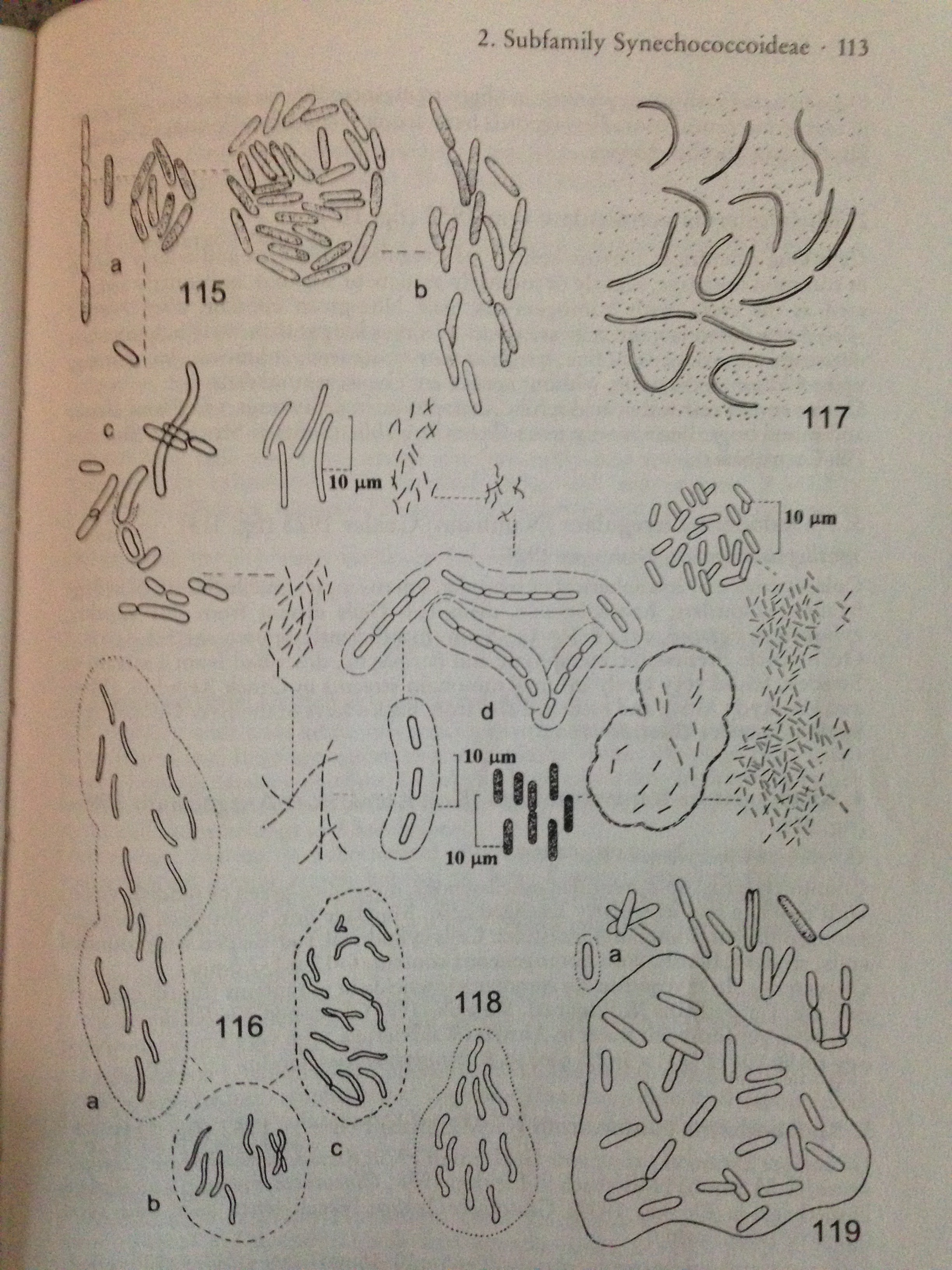
In the picture above, figures 115 and 116 are specifically Rhabdoderma lineare. Now seeing these figures, it is very plausible to see how one can confuse this with other genera such as Synechococcus.
Rhabdoderma lineare is described as:
- Colonies small, few-celled, or later sometimes multicellular , with cells orientated in more or less the same direction.
- Mucilage scarcely visible, fine, colorless, diffluent at the margin.
- Single cells without or sometimes with very five hyaline envelopes.
- Cells long cylindrical, rod-shaped, straight, or slightly arcuate, occasionally in pseudo filamentous rows.
- Size: (3) 4-10 (22) x (0.8) 1.5-3 (3.5) um.
- Cells in colonies sometimes of different lengths, dividing asymmetrically.
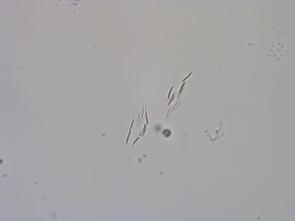
I apologize for the picture below. The individual cells are on different planes, giving some depth perception for the colony. So I tried to get the best image I could to depict the organism and the colony shape. Enjoy!!