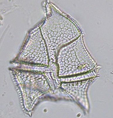
This will be the first of many posts in a collection of what I will call, “Digested Diatoms”. There are many ways to digest diatoms, all of which use some form of chemical reagent in order to remove the organic material. The sole purpose of this is so the minute details of the diatom structures can be clearly seen for taxonomic identification. If you take a look back at some of the previous posts, I point out some of these diatom structures that are pertinent to determine species level identification. One of the common methods is by use of Nitric Acid; so if you have any of that hanging around the house, you can go explore another quarantine hobby!
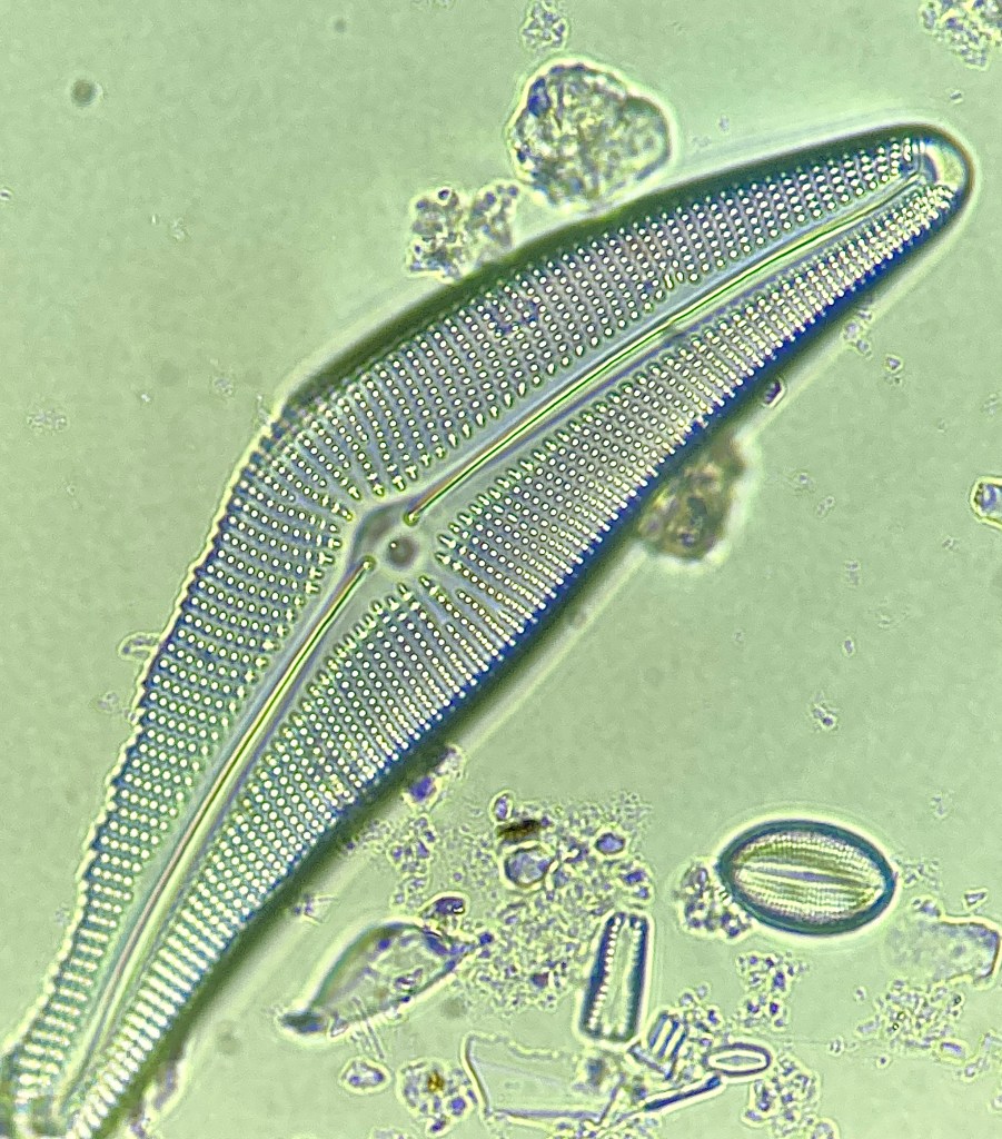
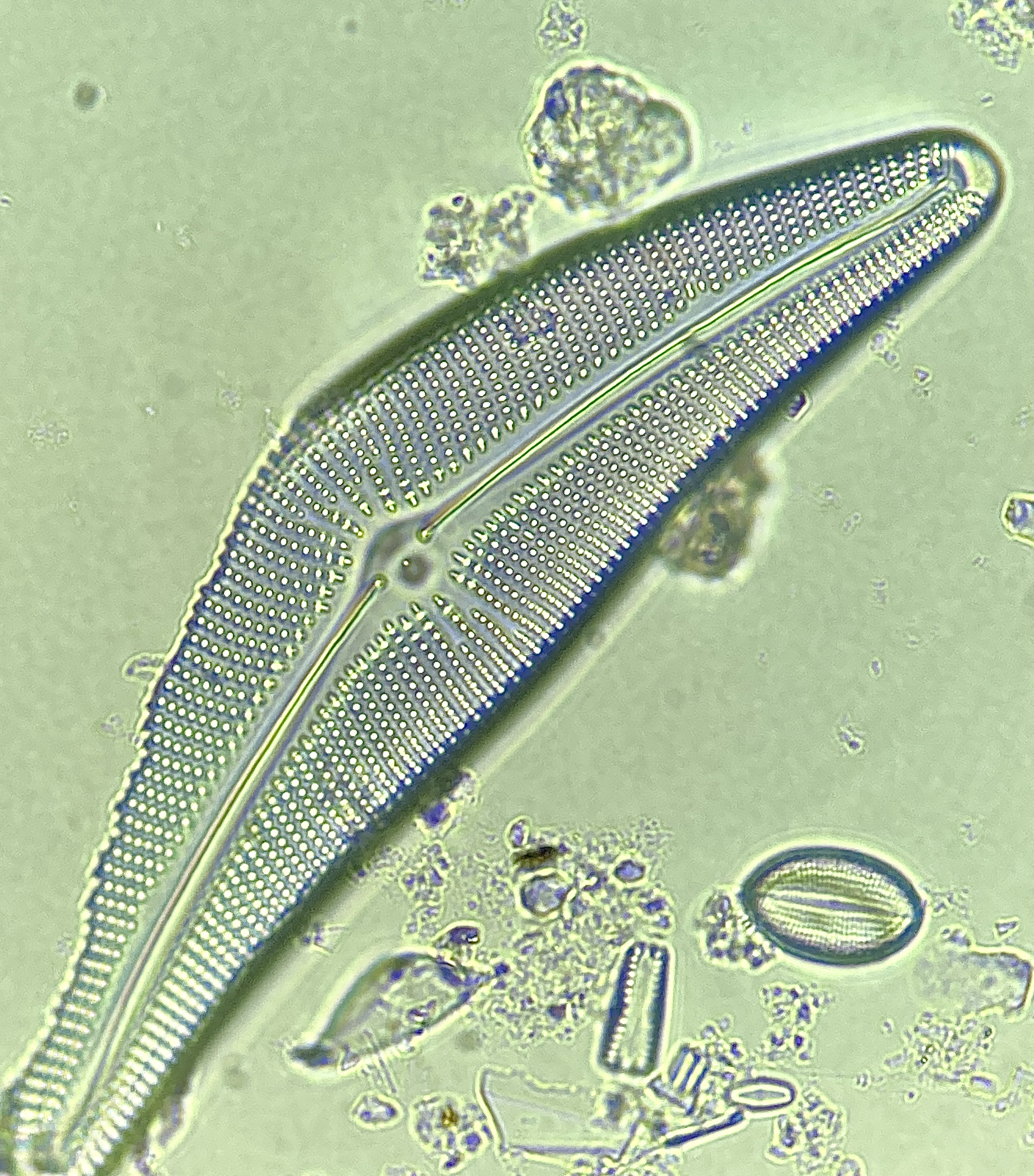
Now, for those of you who are not familiar, this is a diatom in what is called “Valve View”. This is the preferred view to accurately speciate diatoms because the necessary characteristics are in plain view. Its also good to note that digested diatoms make it 1000% easier to speciate because you are viewing the organisms at 100x magnification using immersion oil.
Now, lets take a pause and see what defining characteristics we can see to help us….Take a moment to try for yourselves.
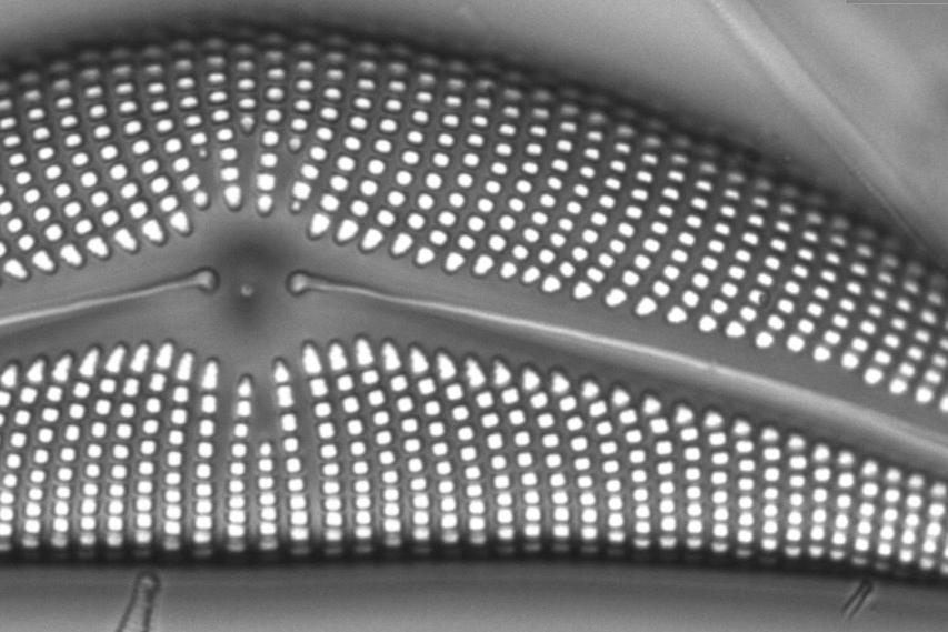
Above is a handy slider application. The Top photo is the one I took, while the bottom photo is a zoomed in SEM image from our friends at Diatoms.org.
The major characteristics we should be focusing on are: Single, Large Stigma is present, 6-8 striae in 10 um in central area (missing scale bar, Sorry!), and Areolae adjacent to axial area elongated (Clearly seen in both pictures).
As always hit the above link to learn more and to see more images provided by Diatoms.org.
If you want to learn more about the specific protocol for cleaning and digesting diatoms, the following link is provided by The Academy of Natural Sciences of Drexel University. This is the specific protocol for NAWQA samples.
If you have any questions/comments, drop a line! Enjoy! and Ill see you soon!
